



Technology
To enhance the standard and capacity of local nuclear medicine services, we continue to stay at the technological forefront by introducing state-of-the-art equipment to Hong Kong. Before service commencement, a series of stringent test will be performed on their new features and safety to optimise the settings and parameters as reference for local clinical usage.

Biograph Vision - SiPM PET/CT Scanner
Our Centre has acquired the first innovative digital SiPM PET/CT scanner in China to provide revolutionary breakthrough in PET imaging accuracy. This innovative technology precisely quantifies cell metabolism according to physiologic characteristics and molecular biochemical properties, which is applied to different clinical specialties such as oncology and neurology. The Nuclear Medicine Specialty team can detect and analyze subtle metabolic changes for underlying pathology. Clinicians can obtain accurate information and prescribe the most suitable treatment regime for patients, such as radiation dosimetry for radiotherapy and radionuclide therapy by oncologist.
- 2 Scanners on 18/F HKSH
- 1 Scanner on 3/F HKSH Eastern Medical Centre
In early 2019, we installed the “Biograph Vision” system with the latest SiPM technology. It has unrivaled hardware specifications and image quality. Its features include:
- Smaller & More Detectors
The dimensions of photon crystal elements have reduced from 4 to 3.2 mm, which allows more detectors per unit area. The number of crystal element has doubled to 60K from the previous generation and the arrays are coped with seamlessly compacted detector. The new assembly significantly enhances image resolution and small lesion detectability. - Enhanced Timing Performance of TOF (Time-of-flight) Hardware
The breakthrough in technology shortens the time resolution of the system, from half to less than 1/4 nanoseconds, and boosts the signal detection sensitivity by 4 times. - Extended Scanning Field
The length has increased from 21 to 26 cm, shortening the total examination time; moreover, bore diameter has expanded to 78 cm (the widest available), sparing more space for body positioning needs. - Multi-parametric PET Imaging
By monitoring PET tracer dynamics inside body instantaneously after injection, the imaging data measures multiple metabolic parameters for absolute quantification of cell metabolism. For example, the absolute metabolic rate and distribution volume in pharmaceutical science calculated by image-based Patlak analysis. - Daily automatic self-calibration
Guarantees image quality and system reliability.

Pro Specta X3 SPECT/CT Scanner
The Symbia Pro Spectra X3 streamlines clinical workflows with automation. It features a multi-slice CT for high-resolution anatomy and various collimators optimised for modern clinical applications, while ensuring fast, high-performance imaging and smooth patient experience.

Biograph mCT Flow PET/CT
Biograph mCT Flow PET/CT scanner is designed with a calm and relaxing ambience. The 135 cm short tunnel can minimize anxiety such as claustrophobia experienced in any other similar examinations, which increases patient’s suitability for the scan. Its 78 cm wide bore also spare more space for patient to maintain scanning posture as well as positioning aids for better comfort.
In view of scan technology, the scanner features patient-specific scanning protocol. The patient bed continuously moves during a scan and personalize imaging parameters according to every patient, which increase the reliability of the examination.

Siemens Intevo SPECT/CT Scanner
Intevo SPECT/CT technology integrates innovative functional SPECT with the structural accuracy of diagnostic multislice CT, enhancing precise localization and nature assessment of diseases, giving new insights into disease processes, and pave the way for new treatment planning and monitoring.

DSPECT Cardiac SPECT Scanner
The DSPECT Cardiac system employs the pioneering solid state detector and semi-conductor crystal technology, making it capable to provide the most advanced electrocardiogram gated cardiac imaging with the highest sensitivity, resolution and rapid acquisition.

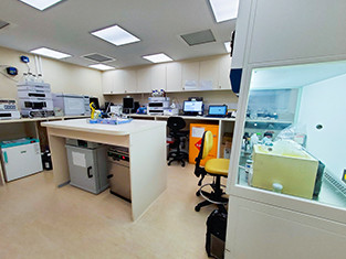
Radiopharmacy Laboratory and Clean Room Facility
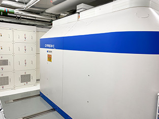
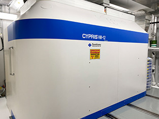
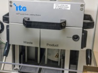
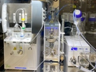
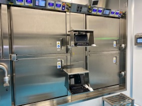
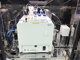
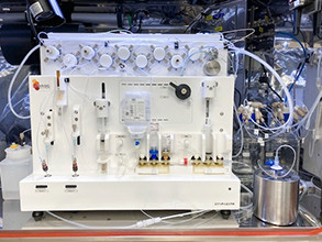
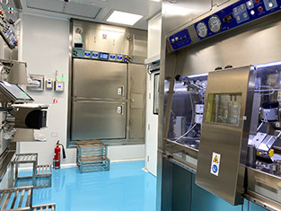
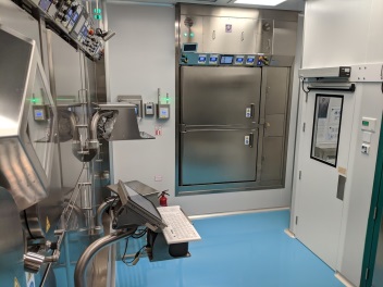
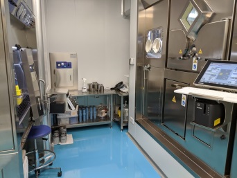
Our latest radiopharmacy laboratory commenced operation in 2020. It is equipped with two medical cyclotrons, clean room and modern radiopharmaceutical synthesizers. The new facility complies with international standard on staff training, equipment and quality check - “Guidelines on Current Good Radiopharmacy Practice (cGRPP) in the Preparation of Radiopharmaceuticals” published by the European Association of Nuclear Medicine (EANM). There are close to 20 PET tracers (labeled with 18-Fluorine, 11-Carbon and 68-Gallium radioisotopes, etc.) currently available for different clinical applications in oncology and neurology.
Medical Cyclotron
Modern Radiopharmaceutical Synthesizers
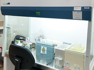
Clean Room
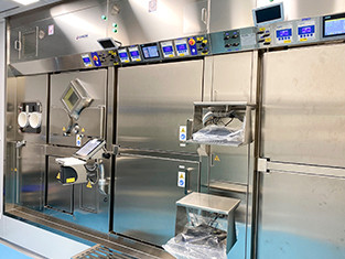
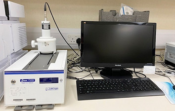
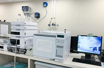
Quality Control Laboratory Settings

uMI Panorama GS - Total-Body PET/CT System
As Hong Kong's first long axial field-of-view (AFOV) Total-Body Positron Emission Tomography/Computed Tomography (PET/CT) scanner, uMI Panorama GS is engineered for high-sensitivity total-body dynamic imaging to optimise clinical workflows while maximising acquisition efficiency and diagnostic precision at a lower radiation dose. In a single scan, the system enables definitive localisation of occult and microscopic primary oncological lesions, thereby enhancing staging accuracy and guiding patient-specific therapeutic regimens.
The system was introduced on 3/F, Li Shu Fong Building, HKSH Eastern Medical Centre in mid-2026. Featuring the world's leading long AFOV digital technology and ultra-sensitivity, it marks a major milestone in both hardware innovations and diagnostic image quality. Key technical and clinical features include:
- Extended Coverage, Shorter Process & Patient comfort
Its 148-cm AFOV enables a single scan from vertex to mid-thigh. By achieving full-body imaging in just 3 to 5 minutes, it can significantly shorten scan time and ensure a simple and smooth process. - Powered by over 410,000 State-of-the-art Digital Crystals & Ultra-High Spatial Resolution
Integrated array of 411,264 ultra-fine 2.76 mm LYSO scintillation crystals coupled with 100% Silicon Photomultiplier (SiPM) digital Modules, the system can achieve an exceptional 2.9mm spatial resolution, markedly enhancing diagnostic sensitivity for hypermetabolic micro-lesions and early-stage malignancies. - 189 Picosecond Time-of-Flight (TOF) Performance
With the breakthrough sub 200 picosecond coincidence timing Resolution, its superior timing resolution substantially elevates the signal-to-noise ratio and contrast, enhancing detection of low-contrast lesions. - Ultra-High Sensitivity & Radiation Dose Optimisation
Its peak system sensitivity of 176 cps/kBq allows ultra-low-dose radiotracer administration and shorter scan durations, fully aligned with radiation protection principles without compromising image quality. - Total-Body Dynamic Multi-Parametric Imaging
Leveraging the 148-cm AFOV, the system captures tracer kinetics immediately post-injection across all major organs (cerebrum, myocardium, liver, lungs, kidneys). It generates comprehensive in vivo pharmacokinetic profiles and absolute metabolic quantification, forming foundational datasets for precision oncology diagnosis and therapy. - AI-Driven Image Reconstruction & Motion Gating
Advanced AI deep learning algorithms inherently capture respiratory motion without external tracking devices, successfully neutralizing respiratory blurring artifacts. Concurrent AI-assisted low-dose CT reconstruction minimizes radiation exposure while preserving structural image quality.

Biograph Vision - PET/CT Simulation for Radiotherapy Treatment Planning
Developed in 2021 PET/CT Simulation is a one-stop imaging facility for both metabolic evaluation and CT simulation in radiotherapy treatment planning. The efficacy of radiotherapy relies on maximizing radiation dose to tumor and preserving normal tissues. With the variety of PET tracers available, PET/CT can delineate the metabolic boundary, quantify aggressiveness and characterize tissue functional status (e.g. cellular differentiation and hypoxia) of targeted tumor.
Our PET/CT scanner with SiPM technology, equipped with an external laser positioning system and a flat indexing pallet, has a precise spatial co-registration and an outstanding image quality. Image data in a single PET/CT scan can streamline the workflow and personalize treatment plan. Biological tumor contours and functional data on PET images can be transferred directly to CT images for dosimetric calculation and dose boosting. It can it be used at pre-treatment stage and within treatment course for interim assessment and adaptive planning.